RESEARCH ARTICLE


Antiproliferative Effects of Ellagic Acid on DU145 Cells


Giuseppe Carota, Giuseppe Sferrazzo, Mariarita Spampinato, Valeria Sorrenti, Luca Vanella*
Department of Drug Science, Biochemistry Section, University of Catania, Viale A. Doria 6, 95125, Catania, Italy


Article Metrics

CrossRef Citations:
4
Total Statistics:

Full-Text HTML Views: 1851
Abstract HTML Views: 868
PDF Downloads: 484
ePub Downloads: 391
Total Views/Downloads: 3594
Unique Statistics:

Full-Text HTML Views: 906
Abstract HTML Views: 454
PDF Downloads: 340
ePub Downloads: 275
Total Views/Downloads: 1975


Creative Commons License
© 2019 Carota et al.

open-access license: This is an open access article distributed under the terms of the Creative Commons Attribution 4.0 International Public License (CC-BY 4.0), a copy of which is available at: https://creativecommons.org/licenses/by/4.0/legalcode. This license permits unrestricted use, distribution, and reproduction in any medium, provided the original author and source are credited.

* Address correspondence to this author at the Department of Drug Science, Biochemistry Section, University of Catania, Viale A. Doria 6, 95125 Catania, Italy; Tel: 0957384077; E-mail: lvanella@unict.it


Abstract

Background:

Prostate Cancer (PC) represents a leading cause of tumor-related death among men in the Western world. Above all, DU145 cell line represents the most particular cells model of PC, derived from a central nervous system metastasis. In recent years, functional and healthy diet has gained a pivotal role in society, allowing the possibility to deal with cancer before its emergence or progression, profiting by anti-tumor properties of dietary phytochemicals. Among them, Ellagic Acid (EA) is found in several fruits and vegetables, whose juice demonstrated antioxidant, anti-carcinogenic and anti-fibrotic properties.

Methods:

DU145 prostate cancer cell line was used to determine the effects of ellagic acid on cell viability. In order to evaluate metastatic feature of DU145, VEGF-A and OPG levels by ELISA assay were assessed. Expression of β-catenin, HO-1, HO-2 and SIRT1, markers of proliferative and defense capacities, were determined by western blotting. To strengthen the study, cell transfection with siRNA β-catenin was performed.

Results:

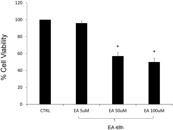
In the presence of EA, the viability of DU145 cells was reduced by about 40 and 50%, respectively after the exposure to 50 and 100 μM concentrations. We also observed a reduction of both levels of VEGF-A and OPG, confirming the important role of EA in facing the metastasis development. EA treatment (50 μM) induced a significant reduction of β-catenin and SIRT1 levels and, similarly, there was a decrease of HO protein expression, more pronounced for HO-2, showing EA activity on the proliferative feature of DU145 cells. Knockdown of β-catenin by siRNA, in the presence of EA treatment, inhibited cell proliferation.

Conclusion:

Ellagic acid exhibits significant antiproliferative effects in our in vitro model of prostate cancer’s metastasis, suggesting that, the use of EA as a multitarget natural compound, may represent a possible strategy for cancer chemoprevention.

Keywords: Antiproliferative, Prostate cancer, Metastasis, DU145e, VEGF-A, OPG.


1. INTRODUCTION

Prostate Cancer (PC) is one of the most common cancer among men and represents a leading cause of tumor-related death, especially in the western world [1-3]. Because of the similarity between PC’s and benign prostate disease’s early symptoms, several patients are treated only in the advanced stage of the tumor progression [4]. Exceeding this point, therapy resistance usually occurs in PC, generating distant metastasis [5]. Patients with high-risk prostate cancer, even in case of radical prostatectomy, present critical conditions due to the presence of delocalized forms of the tumor [6]. During the first steps, genetic alterations and a supporting tumor microenvironment are necessary to confer metastatic status to prostatic cells, permitting displacement toward surrounding tissues [7]. Among all organs compromised by PC’s metastasis, the osteogenic niche makes the bone the most important site of cancer cell proliferation [8, 9]. In fact, a complex cytokine system (RANKL, RANK, OPG) is involved in bone’s resorption and turnover: this seems to be the key point that explains the favorable microenvironment for PC’s cells growing [10]. There are few cell lines available to analyze the metastatic feature of PC and, above all, DU145 cell line represents the most particular cells model that is derived from a central nervous system metastasis, although finding its origin in prostate carcinoma [11, 12]. For this reason, DU145 cells can be chosen to explain the peculiar capacity of PC cells to be invasive and easily capable to move from starting location [13]. The development of PC is a process due to many factors involved in cell's growth and division, thus, inducing a delay or an inhibition of these processes, especially exploiting natural substances commonly present in the diet, could be the right way to prevent cancers from turning clinically significant [14-16]. In recent years, the increasing role of functional and healthy diet allows the possibility to reduce the application of specific drugs that, as cytotoxic agents, are associated to many side-effects that may worsen the quality of patient life [17, 18]. The attention for chemoprevention, thanks to new human acceptance, permits to deal with face cancer before its emergence or progression profiting by anti-tumor properties of dietary phytochemicals, eventually used in combination with treatments of choice [19-21]. Several natural compounds, in particular polyphenols, exhibit an antioxidant activity both in vitro and in vivo [22]. Among them, Ellagic Acid (EA) is found in several fruits and vegetables, like strawberries, blackberries, nuts, and especially in the pomegranates, whose juice demonstrated antioxidant, anti-carcinogenic and anti-fibrotic properties [23-25]. For these reasons, EA has been tested against several cancer types: studied for breast cancer therapy, it inhibited the proliferation and migration of SUM159 and HCC1954 breast cancer cells [26]; as a consequence of p53 activation, it induced growth reduction in colon cancer SW480 cells because of DNA damage [27]; it was also used for bladder cancer, to which it showed an inhibition of extracellular matrix invasion of human bladder cancer cells in response to VEGF-A [28]. The latter, the Vascular Endothelial Growth Factor - A, is the first mediator of normal and pathological angiogenesis, associated with disease progression and recurrence [29]. High levels of VEGF-A are expressed by many tumors, including PC, in which the growth possibility is extended even in metastatic sites.

The aim of this study is to demonstrate the effect of EA treatment on prostate cancer DU145 cell viability and on some specific markers involved in metastatic invasion and migration.

2. MATERIALS AND METHODS

2.1. Cell Culture

DU145 cells (Human prostate carcinoma, epithelial-like cell line) were purchased from American Type Culture Collection (Manassas, VA, USA) and grown in DMEM supplemented with 10% Fetal Bovine Serum (FBS), 0,1% streptomycin-penicillin, 1% L-glutamine and 1% non-essential amino acids. Cells were incubated at 37° C in a 5% CO2 humidified atmosphere and maintained at sub-confluency by passaging with trypsin-EDTA (Gibco, NY, USA).

2.2. Cell Viability Assay

DU145 cells were seeded at a concentration of 2×10⁵ cells per well of a 96-well, flat-bottomed 200-μl microplate. Cells were incubated at 37°C in a 5% CO2 humidified atmosphere and cultured for 48h in the presence and absence of different concentrations (5–100 μM) of EA (≥95% - HPLC – powder, from tree bark) (Sigma-Aldrich. ST Louis. MO-USA). Four hours before the end of the treatment time, 20 μl of 0.5% 3-(4,5-dimethylthiazol-2-yl)-2,5-diphenyltetrazolium bromide (M TT) in Phosphate-Buffered Saline (PBS) was added to each microwell as previously described [30]. The optical density was measured using a microplate spectrophotometer reader (Thermo Labsystems Multiskan, Milano, Italy) at λ=570 nm.

2.3. VEGF-A and OPG Measurements

Cells were seeded at a constant density to obtain identical experimental conditions in the different tests and to achieve a high accuracy of the measurements. VEGF-A and OPG levels were determined in the culture supernatant using an ELISA kit (AssayGate, Ijamsville, MD, USA). The assays were performed according to manufacturer’s guidelines. Results were expressed as pg/mL.

2.4. Immunoblotting of Signaling Proteins

Cells were trypsinized, pelleted by centrifugation, and lysed with lysis buffer supplemented with protease and phosphatase inhibitors (complete™ Mini and PhosSTOP™; Roche Diagnostics, Indianapolis, IA). Protein levels were quantified using a commercial assay (Bio-Rad, Hercules, CA). Protein samples were applied to Sodium Dodecyl Sulfate (SDS) polyacrylamide gel (10%–15%), electrophoresed under denaturing conditions, and electrotransferred onto PVDF Immobilon-P membrane (Amersham Pharmacia, Piscataway, NJ) using a semidry transfer apparatus (Bio-Rad). Membranes were blocked with Odyssey® Blocking Buffer (TBS) (LI-COR, Lincoln, NE) for 1 h at room temperature. Primary antibodies (1:500 – 1:1,000 dilution) in blocking buffer supplemented with 0.1% Tween 20 (Fisher Scientific) were incubated overnight at 4°C, washed in Tris-Buffered Saline (TBS) supplemented with 0.1% Tween 20, and then incubated with the appropriate fluorophore-conjugated secondary antibodies (1:5000–1:20000) (LI-COR). Detection and quantification of signals were completed with a LI-COR Odyssey instrument (LI-COR).

2.5. siRNA Transfection

Transfection was carried out using a solution mix composed of transfection reagent and β-catenin siRNA diluted in siRNA transfection medium (Santa Cruz Biotechnology). DU145 cells were incubated with the solution mix for 5h and then DMEM supplemented with 20% FBS and 1% penicillin /streptomycin was added for the next 24h. After incubation, the cells were treated with EA (50μM) for 48h. A non-targeting siRNA solution (Ambion) was used as negative control.

3. RESULTS

3.1. Effect of Ellagic Acid on Cell Viability

The viability of DU145 cells, in the presence of EA, was assessed by the MTT assay, testing three different concentrations (5, 50, 100 μM). After 48h of EA treatment, the lowest examined concentration did not demonstrate a significant cytotoxic effect, comparing with the untreated cells representing the control group. Contrary, as seen in Fig. (1), we observed a noticeable loss of vitality after EA treatment at the highest concentrations. In particular, cell viability was reduced by about 40 and 50% after the exposure respectively to the 50 and 100μM.

Fig. (1). Cell growth inhibition, determined using MTT assay, of DU145 human prostate cancer cells, untreated and treated with EA at different concentrations (5, 50, 100 μM) for 48h. Data are the means ± SD of 4 experiments performed in triplicate. * p < 0.05 versus untreated cells.

3.2. Effect of Ellagic Acid on VEGF-A and OPG

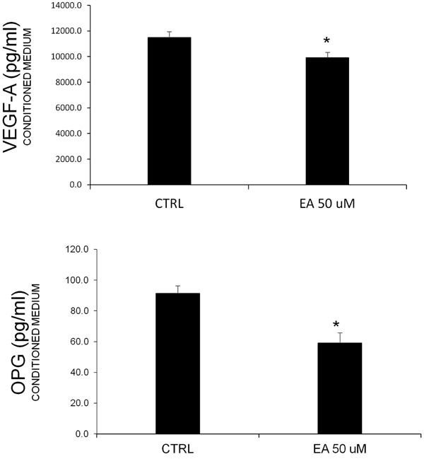
Prostate cancer is known to be associated with bone metastases. Additionally, the involvment of angiogenic factors related to cell proliferation is common. In order to consider these typical features, we measured the VEGF-A and OPG levels in conditioned media obtained after EA treatment (50 μM). The ELISA assay (Fig. 2) showed a decrease of both proteins, confirming the hypothesis about the effect of EA on the release of these factors.

3.3. Effect of Ellagic Acid on β-catenin, HO-1, HO-2 and SIRT1 Levels

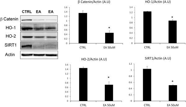
To evaluate the suppression of proliferative and defence capacities of DU145 cells after EA treatment, we assessed the levels of β-catenin, HO-1, HO-2 and SIRT1 (Fig. 3). The densitometric analysis, after normalization with beta-actin, showed that EA treatment, at the concentration previously tested (50 μM), induced a significant reduction of β-catenin and SIRT1 levels. Similarly, we observed a decrease of Heme Oxygenase (HO) protein expression, particularly marked for the constitutive isoform (HO-2).

3.4. Effect of Silencing β-catenin on Cell Proliferation

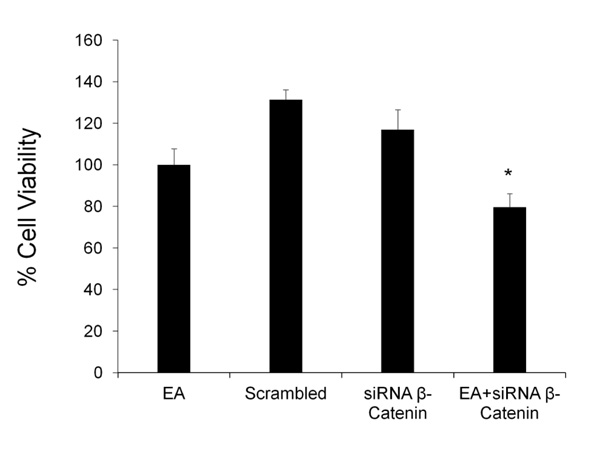
The effect of EA on cell proliferation was tested after silencing β-catenin gene. DU145 cells were treated with EA (50 μM) for 48h in the presence and absence of siRNA against β-catenin; cell viability was detected by MTT assay. Fig. (4) shows that β-catenin siRNA caused a slight decrease in cell viability, while scrambled siRNA did not demonstrate any toxicity. The combination of EA and β-catenin siRNA induced a significant additional reduction of cell viability compared to EA group.

4. DISCUSSION

Prostate cancer is a solid tumor that owns some peculiar features: it is difficult to diagnose in early stages, it shows very high resistance during treatment with chemotherapeutical drugs and, because of its inclination to develop metastasis, the choice of the best drug is complicated and influenced by properties of new affected tissues. This condition leads to patients to embrace new strategies that, in last years, concern chemoprevention and using bioactive compounds. Among natural polyphenols we tested on DU145 PC cells, EA was the best capable compound to face PC’s progression, acting on some specific markers. In the early phase, we assessed EA’s activity on DU145 cells viability, evaluating mitochondrial damage in these cells by MTT assay. Obtained results confirmed our previous study [31], showing a halving of cells viability compared to untreated control, using the highest concentration of EA (50-100 μM). Fig. (1) shows the functional activity of EA stopping cells growth, inhibiting their proliferation. The possible action on mitochondrial function was reported by a similar study, based on EA’s effect on colon cancer Caco-2 cells, that demonstrated a comparable loss of viability on these cells [32]. Although tested in different cell lines and indepen dently of chosen concentrations, EA leads to a halt on cancer cells proliferation, demonstrating the central role of mitochondria in cancer cells survival [33]. Known the EA’s adapt concentration to observe an efficacy on DU145 cells, our analysis continued evaluating resistance and migration features, proper of PC cells [34]. β-catenin is considered part of a protein complex involved in signal transduction that regulates tissues development and biological processes [35, 36]. This pathway is mediated by the Wingless-type (Wnt) proteins, and influences cell apoptosis and tumorigenesis [37, 38]. Wnt protein, binding its cell surface receptor Fz (Frizzled), triggers a signalling cascade through β-catenin, that induces CyclinD1 gene, involved in cell proliferation [39-41]. Is also known the association between β-catenin and the cell adhesion molecule E-cadherin, that represents an essential factor during migration phase of cancer progression [42]. A recent study reported the efficacy of quercitin, another natural compound, on β-catenin signal, whose reduction is associated with the arrest of carcinogenic process, leading to an antiproliferative and apoptotic effect on colon cancer cell lines [43, 44]. Similarly, our results demonstrate a strong reduction of β-catenin protein expression, suggesting that EA could reduce DU145 cells proliferation inhibiting cancer progression. We next investigated whether silencing β-catenin could affect the reduction of cell proliferation mediated by EA. As shown in Fig. (4), and according to previously published results [45-47] pre-treatment with siRNA β-catenin enhanced anti-proliferative effect of EA. SIRT-1 is a member of the sirtuin family of histone deacetylase that is nicotinamide adenine dinucleotide (NAD+)-dependent, and is involved in aging and in cancer growth [48, 49]. The role of SIRT1 as oncogenic protein is explained by the list of its substrates, inhibited by deacetylation, whose function as tumor suppressors is inactivated: Bax, Ku70, FOXO, the Retinoblastoma (Rb) protein, and p53 [50-52]. It has been reported that SIRT1 is significantly overexpressed in many types of cancer, including colon cancer, acute myeloid leukemia and prostate cancer [53-55]. Our results suggest that EA could play a significant role in the reduction of DU145 cells growth and viability even through SIRT1 inhibition, permitting to onco-suppressor genes to induce apoptosis in cancer cells. Many cancers, including PC, overexpress their cytoprotection system leading to stronger resistance to physiological defence or drugs effects. Several studies reported that Heme Oxygenase (HO) system seems to play a major role in cancer cell protection, guaranteeing regulation for redox homeostasis [56-60]. Normal and tumor cells own two different isoforms of HO: one of them (HO-1), is inducible by oxidative stress or inflammatory conditions [61]; the other one (HO-2) represents the constitutive isoform, contributing to basal physiological functions including a decrease in oxidative stress, inflammation, and protection against apoptosis. The main role of HO is to catalyse the rate-limiting step in heme degradation, causing the production of Carbon Monoxide (CO), iron and biliverdin; besides that HO system has been also involved in the development of drug-resistance in various tumors [62-65]. After EA treatment, we observed a strong reduction of HO levels which it is more pronounced for constitutive isoform, showing high capacities of EA to decrease the basal defence features of PC cells, in which progression is known HO involvedness [66]. Inhibiting overexpression of both inducible and constitutive isoforms of HO and the previously mentioned SIRT1, EA demonstrated its contribution to decrease cytoprotective system in DU145 cells, perhaps activating apoptotic pathways that could stop PC cells growth [67]. Every type of cancer cells, including PC cells, needs to develop new vascular structures in surrounding tissues, to guarantee their own survival. This serious condition shows an invasive step of cancer growth, especially associated with metastasis, that permits cancer cells to become part of new tissue, fitting in different location. VEGF-A is the most important factor involved in cancer angiogenesis, stimulating endothelial cells proliferation and formation of new capillary vessels tumor-related [68-71]. In Fig. (2), we observed a statistically significant decrease of VEGF-A level in presence of EA, confirming our previous results obtained on PC LnCap cells [72]. The efficacy of EA on DU145 cells was also demonstrated evaluating the release of Osteoprotegerin (OPG), a peculiar marker involved in bone metastasis. In healthy bone, osteoblastic cells play a central role in bone turnover through the regulation of osteoclastogenic activity [73]. This microenvironment is modified in the presence of PC cells, producing specific effectors that allow an unchecked growth [74]. One of them, OPG, inhibits osteoclastogenesis, avoiding bone remodeling and permitting bone cells to proliferate [75, 76]. It has been reported that PC cells release specific exosomes, which content is transferred to target cells [77]. Different miRNAs, contained in these vesicles, seem to promote tumor metastasis [78] and, one in particular (miR-141-3p), is transferred to osteoblasts promoting their activity, leading to the formation of a bone-metastasis microenvironment. Interestingly, results show an increased OPG release by osteoblast activity, confirming their importance in tumor progression [79]. An altered balance between osteoblast and osteoclast promotes metastatic potential in human prostate cancer [80]. Reduction of OPG levels in DU145 cells culture medium after treatment with EA, demonstrates the role of EA in restoring bone microenvironment and exploiting osteoclastic cell activity to reduce tumor growth.

Fig. (2). VEGF-A and OPG levels in DU145 cells untreated and treated for 48h with EA (50 μM). Values represent the means ± SD of 4 experiments performed in triplicate. * Significance versus untreated control cells: p < 0.05.

Fig. (3). Effect of EA (50 μM) on β-catenin, HO-1, HO-2 and SIRT1 expressions in cultured DU145 cells. Results, expressed as arbitrary units (AU), represent the mean ± SD of 4 experiments performed in triplicate. Significance of 50 μM EA versus control; * p < 0.05. Representative Western blotting of β-catenin, HO-1, HO-2 and SIRT1 protein expression in cultured DU145 cells.

Fig. (4). Cell viability of DU145 cells transfected with EA, scramble and β-catenin siRNA was determined by MTT assay. Cells treated with siRNA targeting β-catenin and EA showed a decreased proliferation compared to EA group (* p<0.05).

CONCLUSION

The present study shows the importance of ellagic acid, as a multivalent natural compound, inhibiting DU145 proliferation through different biochemical mechanisms. Our results suggest that EA could reduce main features of PC related to invasiveness, angiogenesis and metastatic dislocation, noticeable from modulation of factors involved in these processes. In conclusion, we demonstrated that EA treatment may represent an innovative strategy for cancer chemoprevention, supporting traditional therapies.

ETHICS APPROVAL AND CONSENT TO PARTICIPATE

Not applicable.

HUMAN AND ANIMAL RIGHTS

No animals/humans were used for studies that are the basis of this research.

CONSENT FOR PUBLICATION

Not applicable.

AVAILABILITY OF DATA AND MATERIALS

Not applicable.

FUNDING

None.

CONFLICT OF INTEREST

The authors declare no conflict of interest, financial or otherwise.

ACKNOWLEDGEMENTS

Declared none.

REFERENCES

[1] Al-Monajjed, R.; Arsov, C.; Albers, P. Prostate cancer screening: controversies and suggested solutions. Bundesgesundheitsblatt Gesundheitsforschung Gesundheitsschutz, 2018, 61(12), 1544-1550.
[2] Ferlay, J.; Shin, H.R.; Bray, F.; Forman, D.; Mathers, C.; Parkin, D.M. Estimates of worldwide burden of cancer in 2008: GLOBOCAN 2008. Int. J. Cancer, 2010, 127(12), 2893-2917.
[3] Ferlay, J.; Steliarova-Foucher, E.; Lortet-Tieulent, J.; Rosso, S.; Coebergh, J.W.; Comber, H.; Forman, D.; Bray, F. Cancer incidence and mortality patterns in Europe: estimates for 40 countries in 2012. Eur. J. Cancer, 2013, 49(6), 1374-1403.
[4] Salinas, C.A.; Tsodikov, A.; Ishak-Howard, M.; Cooney, K.A. Prostate cancer in young men: an important clinical entity. Nat. Rev. Urol., 2014, 11(6), 317-323.
[5] Hu, S.; Li, L.; Huang, W.; Liu, J.; Lan, G.; Yu, S.; Peng, L.; Xie, X.; Yang, L.; Nian, Y.; Wang, Y. CAV3.1 knockdown suppresses cell proliferation, migration and invasion of prostate cancer cells by inhibiting AKT. Cancer Manag. Res., 2018, 10, 4603-4614.
[6] Sundi, D.; Tosoian, J.J.; Nyame, Y.A.; Alam, R.; Achim, M.; Reichard, C.A.; Li, J.; Wilkins, L.; Schwen, Z.; Han, M. Outcomes of very high-risk prostate cancer after radical prostatectomy: Validation study from 3 centers. Cancer, 2018.
[7] Chung, L.W.; Baseman, A.; Assikis, V.; Zhau, H.E. Molecular insights into prostate cancer progression: the missing link of tumor microenvironment. J. Urol., 2005, 173(1), 10-20.
[8] Wang, H.; Tian, L.; Liu, J.; Goldstein, A.; Bado, I.; Zhang, W.; Arenkiel, BR; Li, Z.; Yang, M.; Du, S. The osteogenic niche is a calcium reservoir of bone micrometastases and confers unexpected therapeutic vulnerability. Cancer cell, 2018, 34(5), 823-839 e827.
[9] Wang, N; Docherty, FE; Brown, HK; Reeves, KJ; Fowles, AC; Ottewell, PD; Dear, TN; Holen, I; Croucher, PI; Eaton, CL Prostate cancer cells preferentially home to osteoblast-rich areas in the early stages of bone metastasis: evidence from in vivo models. JBMR-ASBMR., 2014, 29(12), 2688-2696.
[10] Wada, T.; Nakashima, T.; Hiroshi, N.; Penninger, J.M. RANKL-RANK signaling in osteoclastogenesis and bone disease. Trends Mol. Med., 2006, 12(1), 17-25.
[11] Alimirah, F.; Chen, J.; Basrawala, Z.; Xin, H.; Choubey, D. DU-145 and PC-3 human prostate cancer cell lines express androgen receptor: implications for the androgen receptor functions and regulation. FEBS Lett., 2006, 580(9), 2294-2300.
[12] Stone, K.R.; Mickey, D.D.; Wunderli, H.; Mickey, G.H.; Paulson, D.F. Isolation of a human prostate carcinoma cell line (DU 145). Int. J. Cancer, 1978, 21(3), 274-281.
[13] Pulukuri, S.M.; Gondi, C.S.; Lakka, S.S.; Jutla, A.; Estes, N.; Gujrati, M.; Rao, J.S. RNA interference-directed knockdown of urokinase plasminogen activator and urokinase plasminogen activator receptor inhibits prostate cancer cell invasion, survival, and tumorigenicity in vivo. J. Biol. Chem., 2005, 280(43), 36529-36540.
[14] Naiki-Ito, A.; Chewonarin, T.; Tang, M.; Pitchakarn, P.; Kuno, T.; Ogawa, K.; Asamoto, M.; Shirai, T.; Takahashi, S. Ellagic acid, a component of pomegranate fruit juice, suppresses androgen-dependent prostate carcinogenesis via induction of apoptosis. Prostate, 2015, 75(2), 151-160.
[15] Syed, D.N.; Khan, N.; Afaq, F.; Mukhtar, H. Chemoprevention of prostate cancer through dietary agents: progress and promise. Cancer Epidemiol. Biomarkers Prev., 2007, 16(11), 2193-2203.
[16] Sorrenti, V.; Vanella, L.; Acquaviva, R.; Cardile, V.; Giofrè, S.; Di Giacomo, C. Cyanidin induces apoptosis and differentiation in prostate cancer cells. Int. J. Oncol., 2015, 47(4), 1303-1310.
[17] Onaolapo, A.Y.; Onaolapo, O.J. Nutraceuticals and diet-based phytochemicals in type 2 diabetes mellitus: from whole food to components with defined roles and mechanisms. Curr. Diabetes Rev., 2018.
[18] Braun-Falco, M; Holtmann, C; Lordick, F; Ring, J Follicular drug reaction from cetuximab: a common side effect in the treatment of metastatic colon carcinoma Der Hautarzt; Zeitschrift fur Dermatologie, Venerologie, und verwandte Gebiete, 2006, 57(8), 701-704.
[19] Khan, N.; Afaq, F.; Mukhtar, H. Cancer chemoprevention through dietary antioxidants: progress and promise. Antioxid. Redox Signal., 2008, 10(3), 475-510.
[20] Vanella, L.; Di Giacomo, C.; Acquaviva, R.; Barbagallo, I.; Cardile, V.; Kim, D.H.; Abraham, N.G.; Sorrenti, V. Apoptotic markers in a prostate cancer cell line: effect of ellagic acid. Oncol. Rep., 2013, 30(6), 2804-2810.
[21] Perrone, A.; Capasso, A.; Festa, M.; Kemertelidze, E.; Pizza, C.; Skhirtladze, A.; Piacente, S. Antiproliferative steroidal glycosides from Digitalis ciliata. Fitoterapia, 2012, 83(3), 554-562.
[22] Kang, N.J.; Shin, S.H.; Lee, H.J.; Lee, K.W. Polyphenols as small molecular inhibitors of signaling cascades in carcinogenesis. Pharmacol. Ther., 2011, 130(3), 310-324.
[23] Kaplan, M.; Hayek, T.; Raz, A.; Coleman, R.; Dornfeld, L.; Vaya, J.; Aviram, M. Pomegranate juice supplementation to atherosclerotic mice reduces macrophage lipid peroxidation, cellular cholesterol accumulation and development of atherosclerosis. J. Nutr., 2001, 131(8), 2082-2089.
[24] Han, D.H.; Lee, M.J.; Kim, J.H. Antioxidant and apoptosis-inducing activities of ellagic acid. Anticancer Res., 2006, 26(5A), 3601-3606.
[25] Ceci, C.; Lacal, P.M.; Tentori, L.; De Martino, M.G.; Miano, R.; Graziani, G. Experimental evidence of the antitumor, antimetastatic and antiangiogenic activity of ellagic acid. Nutrients, 2018, 10(11), E1756.
[26] Jaman, M.S.; Sayeed, M.A. Ellagic acid, sulforaphane, and ursolic acid in the prevention and therapy of breast cancer: current evidence and future perspectives. Breast Cancer, 2018, 25(5), 517-528.
[27] Narayanan, B.A.; Re, G.G. IGF-II down regulation associated cell cycle arrest in colon cancer cells exposed to phenolic antioxidant ellagic acid. Anticancer Res., 2001, 21(1A), 359-364.
[28] Ceci, C.; Tentori, L.; Atzori, M.G.; Lacal, P.M.; Bonanno, E.; Scimeca, M.; Cicconi, R.; Mattei, M.; de Martino, M.G.; Vespasiani, G.; Miano, R.; Graziani, G. Ellagic acid inhibits bladder cancer invasiveness and in vivo tumor growth. Nutrients, 2016, 8(11), E744.
[29] Ferrara, N.; Davis-Smyth, T. The biology of vascular endothelial growth factor. Endocr. Rev., 1997, 18(1), 4-25.
[30] Raffaele, M; Barbagallo, I; Licari, M; Carota, G; Sferrazzo, G; Spampinato, M; Sorrenti, V; Vanella, L N-Acetylcysteine (NAC) ameliorates lipid-related metabolic dysfunction in bone marrow stromal cells-derived adipocytes. eCAM, 2018, 2018, 5310961.
[31] Vanella, L.; Barbagallo, I.; Acquaviva, R.; Di Giacomo, C.; Cardile, V.; Abraham, N.G.; Sorrenti, V. Ellagic acid: cytodifferentiating and antiproliferative effects in human prostatic cancer cell lines. Curr. Pharm. Des., 2013, 19(15), 2728-2736.
[32] Larrosa, M.; Tomás-Barberán, F.A.; Espín, J.C. The dietary hydrolysable tannin punicalagin releases ellagic acid that induces apoptosis in human colon adenocarcinoma Caco-2 cells by using the mitochondrial pathway. J. Nutr. Biochem., 2006, 17(9), 611-625.
[33] Skoda, J.; Borankova, K.; Jansson, P.J.; Huang, M.L.; Veselska, R.; Richardson, D.R. Pharmacological targeting of mitochondria in cancer stem cells: An ancient organelle at the crossroad of novel anti-cancer therapies. Pharmacol. Res., 2019, 1(139), 298-313.
[34] Pitchakarn, P.; Chewonarin, T.; Ogawa, K.; Suzuki, S.; Asamoto, M.; Takahashi, S.; Shirai, T.; Limtrakul, P. Ellagic acid inhibits migration and invasion by prostate cancer cell lines. Asian Pac. J. Cancer Prev., 2013, 14(5), 2859-2863.
[35] Polakis, P. Wnt signaling and cancer. Genes Dev., 2000, 14(15), 1837-1851.
[36] Schneider, JA; Logan, SK Revisiting the role of Wnt/beta-catenin signaling in prostate cancer. Molecular and cellular endocrinology., 2018, 462(Pt A), 3-8.
[37] Madan, B.; Virshup, D.M. Targeting Wnts at the source--new mechanisms, new biomarkers, new drugs. Mol. Cancer Ther., 2015, 14(5), 1087-1094.
[38] Rajagopal, C.; Lankadasari, M.B.; Aranjani, J.M.; Harikumar, K.B. Targeting oncogenic transcription factors by polyphenols: A novel approach for cancer therapy. Pharmacol. Res., 2018, 130, 273-291.
[39] Karimaian, A.; Majidinia, M.; Bannazadeh Baghi, H.; Yousefi, B. The crosstalk between Wnt/β-catenin signaling pathway with DNA damage response and oxidative stress: Implications in cancer therapy. DNA Repair (Amst.), 2017, 51, 14-19.
[40] Cianciosi, D.; Varela-Lopez, A.; Forbes-Hernandez, T.Y.; Gasparrini, M.; Afrin, S.; Reboredo-Rodriguez, P.; Zhang, J.; Quiles, J.L.; Nabavi, S.F.; Battino, M.; Giampieri, F. Targeting molecular pathways in cancer stem cells by natural bioactive compounds. Pharmacol. Res., 2018, 135, 150-165.
[41] Barker, N.; Clevers, H. Catenins, Wnt signaling and cancer. BioEssays, 2000, 22(11), 961-965.
[42] Bullions, L.C.; Levine, A.J. The role of beta-catenin in cell adhesion, signal transduction, and cancer. Curr. Opin. Oncol., 1998, 10(1), 81-87.
[43] Refolo, M.G.; D’Alessandro, R.; Malerba, N.; Laezza, C.; Bifulco, M.; Messa, C.; Caruso, M.G.; Notarnicola, M.; Tutino, V. Anti proliferative and pro apoptotic effects of flavonoid quercetin are mediated by CB1 receptor in human colon cancer cell lines. J. Cell. Physiol., 2015, 230(12), 2973-2980.
[44] Park, C.H.; Chang, J.Y.; Hahm, E.R.; Park, S.; Kim, H.K.; Yang, C.H. Quercetin, a potent inhibitor against beta-catenin/Tcf signaling in SW480 colon cancer cells. Biochem. Biophys. Res. Commun., 2005, 328(1), 227-234.
[45] Li, K.; Zhou, Z.Y.; Ji, P.P.; Luo, H.S. Knockdown of β-catenin by siRNA influences proliferation, apoptosis and invasion of the colon cancer cell line SW480. Oncol. Lett., 2016, 11(6), 3896-3900.
[46] Wang, X.H.; Sun, X.; Meng, X.W.; Lv, Z.W.; Du, Y.J.; Zhu, Y.; Chen, J.; Kong, D.X.; Jin, S.Z. beta-catenin siRNA regulation of apoptosis- and angiogenesis-related gene expression in hepatocellular carcinoma cells: potential uses for gene therapy. Oncol. Rep., 2010, 24(4), 1093-1099.
[47] Pu, P.; Zhang, Z.; Kang, C.; Jiang, R.; Jia, Z.; Wang, G.; Jiang, H. Downregulation of Wnt2 and beta-catenin by siRNA suppresses malignant glioma cell growth. Cancer Gene Ther., 2009, 16(4), 351-361.
[48] Jung-Hynes, B.; Nihal, M.; Zhong, W.; Ahmad, N. Role of sirtuin histone deacetylase SIRT1 in prostate cancer. A target for prostate cancer management via its inhibition? J. Biol. Chem., 2009, 284(6), 3823-3832.
[49] Powell, M.J.; Casimiro, M.C.; Cordon-Cardo, C.; He, X.; Yeow, W.S.; Wang, C.; McCue, P.A.; McBurney, M.W.; Pestell, R.G. Disruption of a Sirt1-dependent autophagy checkpoint in the prostate results in prostatic intraepithelial neoplasia lesion formation. Cancer Res., 2011, 71(3), 964-975.
[50] Ota, H.; Tokunaga, E.; Chang, K.; Hikasa, M.; Iijima, K.; Eto, M.; Kozaki, K.; Akishita, M.; Ouchi, Y.; Kaneki, M. Sirt1 inhibitor, Sirtinol, induces senescence-like growth arrest with attenuated Ras-MAPK signaling in human cancer cells. Oncogene, 2006, 25(2), 176-185.
[51] Ford, J.; Jiang, M.; Milner, J. Cancer-specific functions of SIRT1 enable human epithelial cancer cell growth and survival. Cancer Res., 2005, 65(22), 10457-10463.
[52] Chen, H.C.; Jeng, Y.M.; Yuan, R.H.; Hsu, H.C.; Chen, Y.L. SIRT1 promotes tumorigenesis and resistance to chemotherapy in hepatocellular carcinoma and its expression predicts poor prognosis. Ann. Surg. Oncol., 2012, 19(6), 2011-2019.
[53] Jang, K.Y.; Hwang, S.H.; Kwon, K.S.; Kim, K.R.; Choi, H.N.; Lee, N.R.; Kwak, J.Y.; Park, B.H.; Park, H.S.; Chung, M.J.; Kang, M.J.; Lee, D.G.; Kim, H.S.; Shim, H.; Moon, W.S. SIRT1 expression is associated with poor prognosis of diffuse large B-cell lymphoma. Am. J. Surg. Pathol., 2008, 32(10), 1523-1531.
[54] Tseng, R.C.; Lee, C.C.; Hsu, H.S.; Tzao, C.; Wang, Y.C. Distinct HIC1-SIRT1-p53 loop deregulation in lung squamous carcinoma and adenocarcinoma patients. Neoplasia, 2009, 11(8), 763-770.
[55] Huffman, D.M.; Grizzle, W.E.; Bamman, M.M.; Kim, J.S.; Eltoum, I.A.; Elgavish, A.; Nagy, T.R. SIRT1 is significantly elevated in mouse and human prostate cancer. Cancer Res., 2007, 67(14), 6612-6618.
[56] Barbagallo, I.; Giallongo, C.; Volti, G.L.; Distefano, A.; Camiolo, G.; Raffaele, M.; Salerno, L.; Pittala, V.; Sorrenti, V.; Avola, R. Heme oxygenase inhibition sensitizes neuroblastoma cells to carfilzomib. Mol. Neurobiol., 2018, 56(2), 1451-1460.
[57] Li Volti, G.; Tibullo, D.; Vanella, L.; Giallongo, C.; Di Raimondo, F.; Forte, S.; Di Rosa, M.; Signorelli, S.S.; Barbagallo, I. The Heme Oxygenase System in Hematological Malignancies. Antioxid. Redox Signal., 2017, 27(6), 363-377.
[58] Jozkowicz, A.; Was, H.; Dulak, J. Heme oxygenase-1 in tumors: is it a false friend? Antioxid. Redox Signal., 2007, 9(12), 2099-2117.
[59] Vanella, L.; Russo, G.I.; Cimino, S.; Fragala, E.; Favilla, V.; Li Volti, G.; Barbagallo, I.; Sorrenti, V.; Morgia, G. Correlation between lipid profile and heme oxygenase system in patients with benign prostatic hyperplasia. Urology, 2014, 83(6), 1444 e1447-1413.
[60] Waldman, M.; Bellner, L.; Vanella, L.; Schragenheim, J.; Sodhi, K.; Singh, S.P.; Lin, D.; Lakhkar, A.; Li, J.; Hochhauser, E.; Arad, M.; Darzynkiewicz, Z.; Kappas, A.; Abraham, N.G. Epoxyeicosatrienoic acids regulate adipocyte differentiation of mouse 3T3 cells, via PGC-1α activation, which is required for HO-1 expression and increased mitochondrial function. Stem Cells Dev., 2016, 25(14), 1084-1094.
[61] Tibullo, D.; Barbagallo, I.; Giallongo, C.; Vanella, L.; Conticello, C.; Romano, A.; Saccone, S.; Godos, J.; Di Raimondo, F.; Li Volti, G. Heme oxygenase-1 nuclear translocation regulates bortezomibinduced cytotoxicity and mediates genomic instability in myeloma cells. Oncotarget, 2016, 7(20), 28868-28880.
[62] Vanella, L.; Barbagallo, I.; Tibullo, D.; Forte, S.; Zappalà, A.; Li Volti, G. The non-canonical functions of the heme oxygenases. Oncotarget, 2016, 7(42), 69075-69086.
[63] Maines, M.D.; Abrahamsson, P.A. Expression of heme oxygenase-1 (HSP32) in human prostate: normal, hyperplastic, and tumor tissue distribution. Urology, 1996, 47(5), 727-733.
[64] Barbagallo, I.; Parenti, R.; Zappalà, A.; Vanella, L.; Tibullo, D.; Pepe, F.; Onni, T.; Li Volti, G. Combined inhibition of Hsp90 and heme oxygenase-1 induces apoptosis and endoplasmic reticulum stress in melanoma. Acta Histochem., 2015, 117(8), 705-711.
[65] Abraham, N.G.; Junge, J.M.; Drummond, G.S. Translational significance of heme oxygenase in obesity and metabolic syndrome. Trends Pharmacol. Sci., 2016, 37(1), 17-36.
[66] Alaoui-Jamali, M.A.; Bismar, T.A.; Gupta, A.; Szarek, W.A.; Su, J.; Song, W.; Xu, Y.; Xu, B.; Liu, G.; Vlahakis, J.Z.; Roman, G.; Jiao, J.; Schipper, H.M. A novel experimental heme oxygenase-1-targeted therapy for hormone-refractory prostate cancer. Cancer Res., 2009, 69(20), 8017-8024.
[67] He, J.Z.; Ho, J.J.; Gingerich, S.; Courtman, D.W.; Marsden, P.A.; Ward, M.E. Enhanced translation of heme oxygenase-2 preserves human endothelial cell viability during hypoxia. J. Biol. Chem., 2010, 285(13), 9452-9461.
[68] Pepper, M.S.; Ferrara, N.; Orci, L.; Montesano, R. Potent synergism between vascular endothelial growth factor and basic fibroblast growth factor in the induction of angiogenesis in vitro. Biochem. Biophys. Res. Commun., 1992, 189(2), 824-831.
[69] Aldebasi, Y.H.; Rahmani, A.H.; Khan, A.A.; Aly, S.M. The effect of vascular endothelial growth factor in the progression of bladder cancer and diabetic retinopathy. Int. J. Clin. Exp. Med., 2013, 6(4), 239-251.
[70] Pinto, A.; Redondo, A.; Zamora, P.; Castelo, B.; Espinosa, E. Angiogenesis as a therapeutic target in urothelial carcinoma. Anticancer Drugs, 2010, 21(10), 890-896.
[71] Malaponte, G.; Signorelli, S.S.; Bevelacqua, V.; Polesel, J.; Taborelli, M.; Guarneri, C.; Fenga, C.; Umezawa, K.; Libra, M. Increased levels of NF-kB-dependent markers in cancer-associated deep venous thrombosis. PLoS One, 2015, 10(7), e0132496.
[72] Vanella, L.; Di Giacomo, C.; Acquaviva, R.; Barbagallo, I.; Li Volti, G.; Cardile, V.; Abraham, N.G.; Sorrenti, V. Effects of ellagic Acid on angiogenic factors in prostate cancer cells. Cancers (Basel), 2013, 5(2), 726-738.
[73] Sottnik, J.L.; Keller, E.T. Understanding and targeting osteoclastic activity in prostate cancer bone metastases. Curr. Mol. Med., 2013, 13(4), 626-639.
[74] Sottnik, J.L.; Dai, J.; Zhang, H.; Campbell, B.; Keller, E.T. Tumor-induced pressure in the bone microenvironment causes osteocytes to promote the growth of prostate cancer bone metastases. Cancer Res., 2015, 75(11), 2151-2158.
[75] Lynch, C.C.; Hikosaka, A.; Acuff, H.B.; Martin, M.D.; Kawai, N.; Singh, R.K.; Vargo-Gogola, T.C.; Begtrup, J.L.; Peterson, T.E.; Fingleton, B.; Shirai, T.; Matrisian, L.M.; Futakuchi, M. MMP-7 promotes prostate cancer-induced osteolysis via the solubilization of RANKL. Cancer Cell, 2005, 7(5), 485-496.
[76] Sisay, M.; Mengistu, G.; Edessa, D. The RANK/RANKL/OPG system in tumorigenesis and metastasis of cancer stem cell: potential targets for anticancer therapy. Onco Targets Ther., 2017, 10, 3801-3810.
[77] Vlassov, A.V.; Magdaleno, S.; Setterquist, R.; Conrad, R. Exosomes: current knowledge of their composition, biological functions, and diagnostic and therapeutic potentials. Biochim. Biophys. Acta, 2012, 1820(7), 940-948.
[78] Hannafon, B.N.; Ding, W.Q. Intercellular communication by exosome-derived microRNAs in cancer. Int. J. Mol. Sci., 2013, 14(7), 14240-14269.
[79] Ye, Y.; Li, S.L.; Ma, Y.Y.; Diao, Y.J.; Yang, L.; Su, M.Q.; Li, Z.; Ji, Y.; Wang, J.; Lei, L.; Fan, W.X.; Li, L.X.; Xu, Y.; Hao, X.K. Exosomal miR-141-3p regulates osteoblast activity to promote the osteoblastic metastasis of prostate cancer. Oncotarget, 2017, 8(55), 94834-94849.
[80] Karlsson, T.; Sundar, R.; Widmark, A.; Landström, M.; Persson, E. Osteoblast-derived factors promote metastatic potential in human prostate cancer cells, in part via non-canonical transforming growth factor β (TGFβ) signaling. Prostate, 2018, 78(6), 446-456.