All published articles of this journal are available on ScienceDirect.

New Aspects of N-stearoylethanolamine's Neuroprotective Actions in Ageing Rats
 iD
Tetiana Horid’ko1
iD
Andrii Berdyshev1
iD
Olena Meged1
iD
Vitalii Klimashevskyi1
iD
Sergiy Mykhalskyi2
iD
Tetiana Kvitnitskaya-Ryzhova2
iD
Nadiya Hula1
iD
Oksana Tkachenko1
iD
Oleksandr Zhukov1
Authors Info & Affiliations
Abstract
Introduction/ Objective
During aging, numerous pathological changes occur in the brain, including changes in its lipid composition. This leads to impaired memory and cognitive function. All existing geriatric drugs cause side effects. N-acylethanolamines (NAE) are a class of biologically active lipids that are part of the body's endocannabinoid system and play an important role in the functioning of the central nervous system.
This study aimed to investigate the effects of NAE18:0 N-stearoylethanolamine (NSE) on memory, exploration activity, neuronal state, and lipid composition, choline, acetylcholine content, and acetylcholinesterase (AChE) activity in the brain and blood plasma in 18-month-old aging rats.
Methods
All animals were tested using a “New object recognition test” and “Hole board test” to assess rats’ memory and exploration activity. Old rats in the experimental group were orally administered a water suspension of NSE for 10 days. Thin-layer chromatography, ELISA, and mass spectrometry were used.
Results
The use of NSE led to changes in the phospholipid composition and cholesterol content in the frontal cortex and hippocampus, AChE activity, and a slight increase in acetylcholine in the hippocampus and frontal cortex, a decrease in the number of hyperchromic and vacuolated forms of neurons in brain structures, as well as improved memory and exploratory activity in aging rats.
Discussion
The neuroprotective effect of NSE in aged rats may be due to its influence on fatty acid amide hydrolase (FAAH) activity, modulation of the PPARγ/NF-κB signaling pathway, activation of PPAR receptors and α7 nicotinic acetylcholine receptors, and the ability of NSE to influence the initial stages of inflammation characteristic of old age. These hypotheses require further research in the future.
Conclusion
Thus, the results of this study suggest that NSE has a pronounced neuroprotective and antiamnestic effect in age-related changes in the brain. The use of NSE in old rats leads to an improvement in cognitive functions, as well as to a decrease in the severity of morphofunctional disorders in brain structures, including a decrease in signs of age-related neuronal degeneration. The data obtained confirm the participation of NSE as a component of the endocannabinoid system in regulating the cholinergic system and highlight the prospects for further studies of this compound in correcting age-related neurodegenerative processes.
These results could be used to develop a new geriatric drug, taking into account the absence of known adverse reactions in NSE.
1. INTRODUCTION
Aging is a universal biological process characterized by the progressive deterioration of the structure and functional state of tissues and organs. Aging is often accompanied by the development of chronic low-grade inflammation, also known as “inflammaging” [1, 2]. This state is a prerequisite for the emergence of the so-called multiple morbidities, that is, a set of various chronic diseases, particularly metabolic, oncological, autoimmune, neurodegenerative, etc [3], which ultimately leads to fatal consequences for the body.
In recent years, lipids have emerged as a focus of research into the causes and mechanisms of aging. Today, it is known that lipids are essential regulators of various biological processes, such as differentiation, apoptosis, transmembrane traffic, etc [4-7].
Brain tissue is the most enriched in lipid compounds among all body tissues, which indicates the important role of lipids in ensuring the structural features of this organ and regulating its functions. Changes in lipid composition play an essential role in the mechanisms of brain aging. The literature describes a decrease in the content of most lipid classes in the human brain after the age of 50 [8], especially cholesterol levels [9] and changes in phospholipid content (increase or decrease in PtdSer, PtdChol, and PtdEtn) [10].
A considerable decrease in the content of phospholipids (up to 20%) was found in the structures of the human brain responsible for the formation of cognitive function and memory (prefrontal cortex, hippocampus) [11]. It has been shown that a decrease in lipid content in lipid rafts can lead to the development of neurodegenerative processes and cognitive impairment [12].
It is known that the endocannabinoid system is a regulator of memory and learning processes and also plays a significant role in the formation of emotions [13]. This system includes a class of minor lipids - N-acylethanolamines (NAEs); among them, there are both endogenous ligands of cannabinoid receptors (CB) (anandamide) and compounds that mimic the effects of cannabinoids without activating CB receptors. NAEs have many unique properties: adaptogenic, membrane-protective, antioxidant, immunomodulatory, anti-inflammatory, anti-allergic, and others [14]. NAEs also exhibit anxiolytic, analgesic, and antidepressant effects, along with regulating sleep, appetite, and blood-brain barrier permeability [15].
In our previous studies, it was found that one of the saturated NAEs, N-Stearoylethanolamine (NSE) reduced the blood-brain barrier permeability and improved the memory of mice injected with lipopolysaccharide or immunized with the extracellular domain of the α7 subunit of nicotinic acetylcholine receptors [16]. We have recently shown that NSE, administered to rats prior to scopolamine-induced cognitive impairment, prevents memory impairment in animals [17]. In view of this, we hypothesized that NSE may improve the functional state of the aging brain.
This study aimed to investigate the effects of NAE18:0 N-stearoylethanolamine (NSE) on memory, exploration activity, neuronal state, and the lipid composition, choline, acetylcholine content, and acetylcholinesterase (AChE) activity in the brain and blood plasma in aging 18-month-old rats.
2. MATERIALS AND METHODS
2.1. Animals
This study was performed on male Sprague-Dawley rats (n=30) aged 18 months (n=20; body weighs 470-620 g) and 4 months (n=10; body weight 180-278 g). The animals were purchased from the Ukrainian laboratory animal breeding facility “DALI 2001”. The 18-month-old rats were randomly divided into experimental and control groups (n = 10 in each group) using our own script (php 8.3.7, function rand (1,20)). The optimal number of animals in the group was calculated using the computer program “Java applets for power and sample size” [18].
Male rats were selected for this study due to their hormonal stability. Unlike females, males do not undergo cyclical hormonal fluctuations associated with the estrous cycle, resulting in a more stable and predictable endocrine baseline. This absence of estrous variability minimizes scatter in physiological and biochemical responses (e.g., to drug treatment or stress), thereby increasing data reproducibility and reliability.
All rats were kept in the animal facility of Palladin Institute of Biochemistry NAS of Ukraine under standard vivarium conditions (temperature +22 ± 2°C, air humidity 50–60%, 12-h light/dark cycle) in plastic cages (of 5 animals) with iron lattice, with free access to water and food (rodent chow, Animal Feed Manufacturer “Agrovita”, Ukraine). Animals’ health was monitored by weight (weekly), food and water intake, and general assessment of animal activity and fur condition. All experiments involving animals were conducted following established bioethical principles and international regulations for the use of laboratory animals, including EU Directive 2010/63/EU, the European Convention for the Protection of Vertebrate Animals Used for Experimental and Other Scientific Purposes (Strasbourg, 1986), and the Ukrainian Guidelines for Bioethical Assessment of Preclinical and Other Scientific Research on Animals (Kyiv, 2006). The study was approved by the Animal Care and Use Committee of the Palladin Institute of Biochemistry of the NAS of Ukraine (protocol No. 8, 05.05.2021).
Old rats from the experimental group received a 0.5 mL water suspension of NSE orally at a dose of 50 mg/kg body weight daily for 10 days.
Healthy 4-month-old rats did not need treatment, so they were administered distilled water orally instead of NSE. Rats in the control group, aged 8 months, were also administered distilled water orally instead of NSE.
The dose of NSE was selected as the optimal experimental dose for this study based on previous work by Schmid et al., who reported that this concentration of NSE was found during a dog coronary artery occlusion experiment [19]. A water suspension of NSE was prepared by means of an ultrasonic homogenizer UP50H, 50 W, 30 kHz.
N-Stearoylethanolamine was synthesized in the Department of Lipid Biochemistry at Palladin Institute of Biochemistry by the method which consists in the condensation of ethanolamine and stearic acid in an argon atmosphere and at a certain temperature regime as described previously [20]. The purification of the obtained NSE was carried out by recrystallization in ethanol. The final product was identified as a single spot corresponding to the NSE standard (S8439 from Sigma-Aldrich) by thin-layer chromatography in the solvent system chloroform: methanol: 25% ammonia (80: 20: 2).
2.2. Cognitive and Memory Assessments
The “Novel object recognition” (NOR) test and “Hole board test” were performed to assess rats’ memory and exploration activity. The NOR test measures memory by observing whether an animal explores a new object more than a familiar one. The results of the NOR are presented as the Discrimination Index (DI), calculated as the difference in the number of “novel” and “famous” object explorations divided by the total number of explorations [21]. The “Hole board test” assesses novelty-seeking and exploratory activity of the animals and can also be used to study anxiety and cognition by observing head-dipping and nose-poking in holes on a board [22, 23]. The frequency of head-dipping indicates the level of exploration activity in animals. Therefore, the results of the “Hole board test” are presented as the number of times the animal dips its head into the holes.
The ”Hole board test” was conducted on the first day, and the NOR test on the first (habituation phase) and second (training and test phases) days using a wooden box (90 cm x 80 cm x 65cm) with 9 evenly spaced holes. The rat was removed from its home cage and placed in the middle of the open arena with holes to explore freely for 5 minutes. The results of the “Hole board test” are presented as the number of times the animal dips its head into the holes.
On the first NOR test day (habituation phase), the rat was removed from its home cage and placed in the middle of the open arena to explore freely for 5 minutes. The next day, we placed 2 identical objects in opposite quadrants of the open arena. A wooden box was used as an open arena. Then the rat was removed from its home cage and placed in the center of the open arena for free exploration for 10 min (training phase). At the end of the training phase, the rat was removed from the open area and placed in the holding cage for 20 min. After the retention interval, the rat was returned to the open arena with two objects: one “famous” and the other “novel”, for free exploration for 10 minutes (test phase). The number of explorations of the “novel” and “famous” objects was calculated. The results of NOR are presented as the Discrimination Index (DI), calculated as the difference in the number of “novel” and “famous” object explorations divided by the total number of explorations.
All tests were recorded using a video camera (model: A4Tech PK-335MB, software: Amcap-DirectShow Video Capture Sample v.9.00, ©Microsoft Corp.) and analyzed by two independent people who were completely unaware of the animals' group assignment.
2.3. Biochemical Studies
Animals were decapitated under CO2 anesthesia [24]. Whole blood samples and brain areas were isolated within 1- 3 minutes at 4 °C. The blood plasma and separated brain structures were stored at −80 °C for biochemical studies.
2.4. Lipid Analysis
Lipid extraction from rats' hippocampus and frontal cortex tissues was performed using the Bligh and Dyer method [25]. To 75-100 mg of brain tissue, 0.9% NaCl solution was added to form a 10% homogenate. Then, the extraction mixture – chloroform/methanol (1:2) was added to obtain a final solvent ratio of chloroform/methanol/ water (1:2:0.8). After homogenization, chloroform and 0.9% NaCl solution were added to obtain a final solvent ratio - aqueous phase/chloroform/methanol- of 0.9:1:1. The mixture was homogenized and centrifuged for 20 min at 2500 g. The chloroform layer was transferred to a flask and evaporated. Lipid extracts (in 0.5 mL benzene) were stored in a refrigerator at -18° C for lipid analysis.
Phospholipid separation was performed by two-dimensional micro-thin layer chromatography according to the method of Svetashev and Vaskovsky [26, 27] on Sorbfil PTSH-AF-A plates in solvent systems chloroform/methanol/ benzene/28% ammonia in the corresponding volume ratio of 65:30:10:6-8 (1st direction); chloroform/methanol/ benzene/acetone/glacial acetic acid/water in the ratio 70:30:10:5:4:1 (2nd direction).
Diacyl and plasmalogen forms of phospholipids were separated by two-dimensional micro-thin layer chromatography on Sorbfil plates. In the first direction, the separation was carried out in the solvent system of chloroform/methanol/28% ammonia in the ratio 65:35:5. In the second direction, the separation was carried out in 2 stages in different solvent systems: 3M HCl in methanol (stage 1) and chloroform/acetone/methanol/glacial acetic acid/water in the ratio 100:40:20:20:10 (stage 2) [28]. Phospholipid classes were identified as described in [29]. The amount of individual identified phospholipids was estimated by colorimetric measurement of inorganic phosphate (Pi) that was determined spectrophotometrically by the Vaskovsky and Kostecky method [30].
The lipid extract separation into classes carried out by the method of one-dimensional thin-layer chromatography on “Sorbfil” PTSH-P-A plates in the solvent system hexane/diethyl ether/glacial acetic acid (85:15:1). The zone of free cholesterol was removed from the plate, transferred to a microcolumn and eluted with diethyl ether.
Quantitative analysis of free cholesterol was performed using an Agilent 7890-5977 GCMS system equipped with an HP-5MS column (30 m x 250 μm x 0.25 μm) with helium as a carrier gas at 1 ml·min-1 flow rate, and the temperature program was as follows: 60 °С for 1min, then raised to 280 °С at 5 °С·min-1. The final temperature of 280 °C was maintained for 15 minutes. Total time 60 min, injector split 20:1, injected sample volume was 5 μl. Cholesterol content was calculated using a standard curve.
2.5. Determination of Choline, Acetylcholine Content and AChE Activity
The choline and acetylcholine content in the homogenate of the hippocampus and the frontal cortex of the rat brain was determined using the test set “Choline/Acetylcholine Quantification Kit” (Sigma, USA). A set of reagents, the Acetylcholinesterase Activity Assay Kit (Sigma, USA), was used to estimate the activity of AChE in the homogenates of the hippocampus and frontal cortex of rat brains. The detection principle was as follows: AChE catalyzes the hydrolysis of acetylcholine to form choline, and choline reacts with dithio p-nitrobenzoic acid (DTNB) to form 5-mercapto-nitrobenzoic acid (TNB). TNB has an absorption peak at 412 nm. The activity of AChE is calculated by measuring the rate of increase in absorbance at 412 nm. The enzymatic amount that catalyzes the production of 1 nmol TNB by 1 mL of plasma (or by 1 mg of protein of the frontal cortex and hippocampus) per minute is defined as 1 unit. A selection of reagents “Cholinesterase AXX REF HP027.01” (“Filisit-Diagnostika”, Ukraine, 49051 Dnipro, str. Kashtanova, 32) was used to estimate the activity of cholinesterase (ChE) for appropriate blood plasma analysis.
2.6. Morphological Studies
For the morphological studies, the part of the brain of rats that contained the CA1 hippocampal area and sensorimotor part of the frontal cortex was isolated and fixed in 10% neutral buffered formalin, passed through ethanol solutions of increasing concentration, and xylene to paraffin blocks (paraplast type 6, Richard-Allan Scientific, USA) according to the standard method. The rat brain tissue histological sections were stained using Nissl's method with toluidine blue and cresyl violet.
The obtained histological preparations were studied and photographed at x400 magnification using the Olympus BX51 microscope image analysis system (Japan) with Olympus DP-Soft 3.2 software. The numerical density of neurons was calculated as the sum of neurons per test area of 320x240 μm2, expressed in the number of cells per mm2. The neurons were counted on approximately 18-20 test visual fields on preparations (histological sections) from one animal. In each field (area), the cells were counted twice; if the numbers of the counting results did not differ, then no additional counting was done; if the number differed by 1-2 cells, then the arithmetic mean was taken. When counting neurons, in addition to the total number, the number of “unchanged”, “hyperchromic,” and “vacuolated” neurons was determined.
The latter were defined as clearly destructively and dystrophically changed, edematous. Hyperchromic neurons represented a heterogeneous population, comprising both activated cells with intense Nissl staining and a euchromatic nucleus, and destructively altered cells featuring indistinct cytoplasmic organelles and a pyknotic (or hyperchromatic) nucleus. We did not observe reliable differences in the total number of neurons between the experimental groups. However, changes in the redistribution of the ratio of the conditional “unchanged” (normal), “hyperchromic,” and “vacuolated” neurons isolated by us were detected, which is presented in this article.
2.7. Statistical Analysis
Statistical analysis was performed using the IBM SPSS Statistics 23 software, with descriptive statistical methods (means, standard deviations, Shapiro-Wilk test) and one-way analysis of variance (ANOVA), followed by a post hoc Tukey honestly significant difference (HSD) test to assess differences in all measurement indices between groups. Data with p<0.05 were considered significant. Correlations were evaluated using Pearson's correlation test with the MS Excel® function CORREL().
3. RESULTS
3.1. Cognitive and memory assessments
Fig. (1a) demonstrates the DI values in the NOR, and Fig. (1b) – the number of explored board holes (head dipping and nose poking into holes) in the “Hole-board test”.
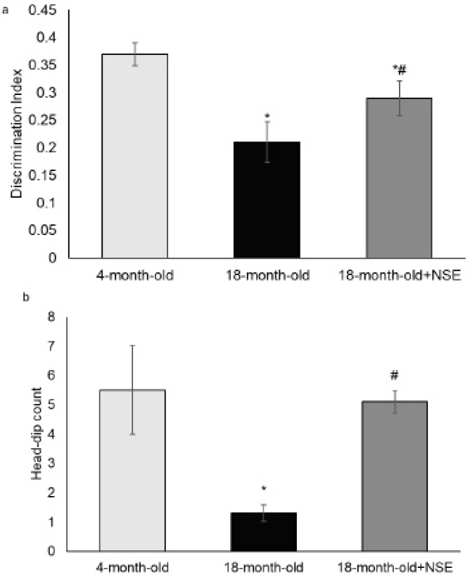
DI (a) and number of head dipping (b). NSE, N-stearoylethanolamine; +NSE, treatment with water suspension of N-stearoylethanolamine at a dose of 50 mg/kg of body weight daily for 10 days; Data expressed as Mean ± SD. The Shapiro-Wilk test was used to assess the normality of the data. One-way ANOVA followed by a post hoc Tukey honestly significant difference (HSD) test was used to assess differences in all measurement indices between groups; *represents a statistically significant difference between 4-month-old and 18-month-old rats' groups at the significance level of 0.05; # represents a statistically significant difference between the 18-month-old rats' group and the same age group after NSE administration at the significance level of 0.05
Fig. (1a) shows that the value of the DI in old rats (Fig. 1a 2nd bar) significantly decreased compared to the value in young rats (Fig. 1a 1st bar). That is, old rats preferred to study an already known (old) object or to study a new and old object simultaneously. This observation indicates the deterioration of short-term memory in 18-month-old rats. Under the treatment of NSE, the DI value increased significantly in old rats: these rats more actively explored a new object, which indicates an improvement in their short-term memory (Fig. 1a, 3rd bar). Fig. (1b) shows that in old rats, the average number of head dips (Fig. 1b, 2nd bar) was significantly lower than in young rats (Fig. 1b, 1st bar), which indicates a reduced level of cognitive activity with aging. Following NSE administration in 18-month-old rats, the number of head dips increased significantly (Fig. 1b, 3rd bar), which may indicate enhanced exploratory activity and cognitive function.
3.2. Biochemical Studies
3.2.1. Lipid Composition of the Brain's Frontal Cortex and Hippocampus
Fig. (2) shows data on the content of diacyl and plasmalogen forms of phospholipids in the hippocampus and frontal cortex of the rat brain.
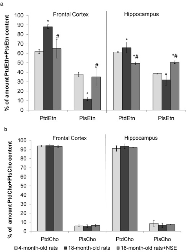
Content of diacyl (a) and plasmalogen (b) forms of phospholipids in the frontal cortex and hippocampus of young and old rats; PtdEtn, phosphatidylethanolamine diacyl form; PlsEtn, phosphatidylethanolamine plasmalogen; PtdCho, phosphatidylcholine diacyl; PlsCho, phosphatidylcholine plasmalogen; NSE, N-stearoylethanolamine; +NSE, treatment with water suspension of N-stearoylethanolamine at a dose of 50 mg/kg of body weight daily for 10 days; Data expressed as Mean ± SD. The Shapiro-Wilk test was used to assess the normality of the data. One-way ANOVA followed by a post hoc Tukey honestly significant difference (HSD) test was used to assess differences in all measurement indices between groups; *, represents a statistically significant difference between 4-month-old (n=10) and 18-month-old (n=10) rat groups at the significance level of 0.05; # represents a statistically significant difference between 18-month-old rats and 18-month-old+NSE group at the significance level of 0.05
Fig. (2a) shows the significant growth of PtdEth and the reduction of PlsEth content in the frontal cortex of old rats compared to young rats. Respective age-induced changes of PtdEth and PlsEth in the hippocampus remained statistically insignificant. At the same time, the content of PtdCho and PlsCho in the old rat group did not change relative to their values in young animals, both in the prefrontal cortex and in the hippocampus (Fig. 2b). Thus, one can see the restoring effect of NSE on PtdEth and PlsEth content in the frontal cortex of aged rats (Fig. 2a), while not affecting the content of PtdCho and PlsCho in the frontal cortex and hippocampus (Fig. 2b).
Fig. (3a, b) demonstrates the content of the major phospholipids in the frontal cortex and hippocampus of rats.
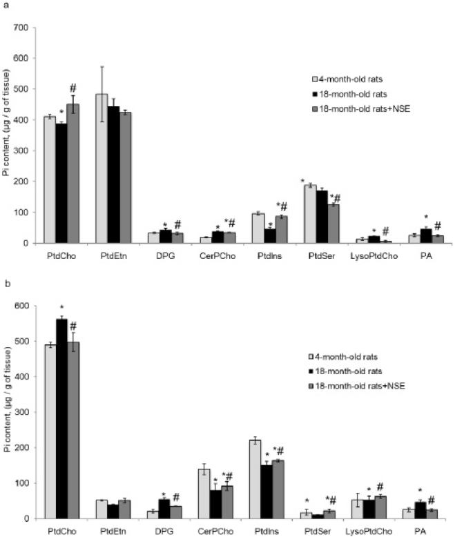
Content of phospholipids (µg/g of tissue) in the frontal cortex (a) and hippocampus (b) of rats; PtdCho, Phosphatidylcholine; PtdEth, Phosphatidylethanolamine; DPG, Diphosphatidylglycerol; CerPCho, Sphingomyelin; PtdIns, Phosphatidylinositol; PtdSer, Phosphatidylserine; LysoPtdCho, Lysophosphatidylcholine; PA, Phosphatidic acid; +NSE, treatment with water suspension of N-stearoylethanolamine at a dose of 50 mg/kg of body weight daily for 10 days; Data expressed as Mean ± SD. The Shapiro-Wilk test was used to assess the normality of the data. One-way ANOVA followed by a post hoc Tukey honestly significant difference (HSD) test was used to assess differences in all measurement indices between groups; *represents a statistically significant difference between 4-month-old (n=10) and 18- month-old rat (n=10) groups at the significance level of 0.05; # represents a statistically significant difference between 18-month-old rats and 18-month-old+NSE group at the significance level of 0.05
Fig. (3) shows that in the hippocampus and frontal cortex of the brain of old rats, there were changes in the content of the main phospholipids compared to the group of young rats. Thus, during aging, the content of PtdCho, Ptdlns, and PtdSer in the frontal cortex and DPG, Ptdlns, and PtdSer in the hippocampus decreased. At the same time, an increase in the levels of DPG, CerPCho, LysoPtdCho, and PA in the frontal cortex and PtdEth and CerPCho in the hippocampus occurred. The administration of NSE to old rats facilitated normalization of the content of DPG and PA in the frontal cortex and PtdCho, LysoPtdCho, DPG, and PA in the hippocampus.
Fig. (4a, b) shows free cholesterol content in the rat frontal cortex and hippocampus.
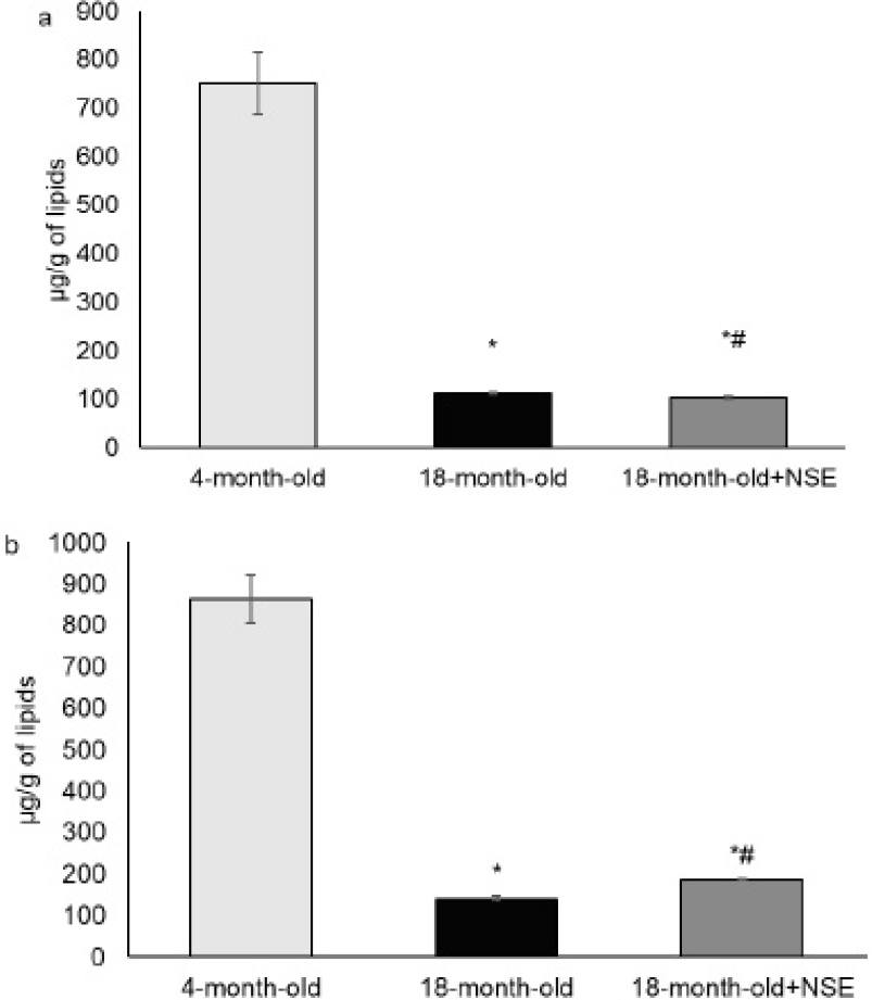
Free cholesterol content in the frontal cortex (a) and hippocampus (b) of 4-month-old and 18-month-old rats; +NSE, treatment with water suspension of N-stearoylethanolamine at a dose of 50 mg/kg of body weight daily for 10 days; Data expressed as Mean ± SD. The Shapiro-Wilk test was used to assess the normality of the data. One-way ANOVA followed by a post hoc Tukey honestly significant difference (HSD) test was used to assess differences in all measurement indices between groups; *represents a statistically significant difference between 4-month-old (n=10) and 18- month-old rat (n=10) groups at the significance level of 0.05; # represents a statistically significant difference between 18-month-old rats and 18-month-old+NSE group at the significance level of 0.05
Fig. (4) shows a decrease in free cholesterol content in the frontal cortex and hippocampus of the rat brain, compared to the levels in young animals. The administration of NSE appears to contribute to an increase in cholesterol content in the hippocampus, while not affecting its amount in the frontal cortex of the brain of old rats.
3.2.2. Choline, Acetylcholine Content and AChE Activity
Fig. (5a-g) presents the results of our experiment, showcasing AChE activity, choline, and acetylcholine content in the hippocampus, frontal cerebral cortex, and blood plasma of rats from different age groups.
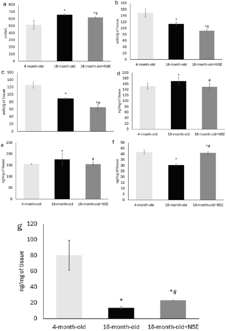
AChE activity in the frontal cortex (a), hippocampus (b), and plasma (c). The enzymatic amount that catalyzes the production of 1 nmol TNB by 1 mL of plasma per minute is defined as 1 unit; Choline content in the frontal cortex (d) and hippocampus (e); Acetylcholine content in the frontal cortex (f) and hippocampus (g) of young and old rats; NSE, N-stearoylethanolamine; +NSE, treatment with water suspension of N-stearoylethanolamine at a dose of 50 mg/kg of body weight daily for 10 days; Data expressed as Mean ± SD. The Shapiro-Wilk test was used to assess the normality of the data. One-way ANOVA followed by a post hoc Tukey honestly significant difference (HSD) test was used to assess differences in all measurement indices between groups; *represents a statistically significant difference between 4-month-old and 18-month-old rat groups at the significance level of 0.05; # represents a statistically significant difference between the 18-month-old rat group and the same age group after NSE administration at the significance level of 0.05
Fig. (5 a, b) demonstrates that in the hippocampus and frontal cortex of old rats' brains, compared with young rats, the activity of AChE is reduced. At the same time, the result showed statistically significant growth of AChE activity in the blood plasma of 18-month-old rats compared to the 4-month-old animals’ group. Administration of NSE to old rats increased the compensatory reduction of AChE activity, which contributes to the restoration of acetylcholine levels both in the hippocampus and in the frontal cortex of the brain. NSE administration positively affected blood plasma AChE activity in old rats, but its level remained higher than in young rats. Fig. (5d, e) demonstrates that the aging rats' frontal cortex and hippocampus contain significantly more choline. Although administering NSE to old rats reduced the level of free choline in the studied brain structures, its content remained significantly higher than in young rats. Fig. (5f, g) shows that aging is accompanied by a significant decrease in the level of acetylcholine in the studied brain structures of rats compared to its level in young animals. NSE administration caused restoration of the content of acetylcholine in the frontal cortex of the brain of 18-month-old rats. In contrast, in the hippocampus of old rats, the content of acetylcholine increased slightly.
3.2.3. Morphological Studies
Fig. (6a, b) illustrates morphological changes in the neurons of the hippocampal CA1 region and sensorimotor part of the frontal cortex of rats during aging.
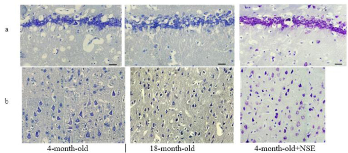
Light microscopy of the neurons of the hippocampal CA1 region (a) and frontal cortex (b). Typical photo of the rat brain tissue histological sections; stained according to Nissl with toluidine blue mixed with cresyl violet; x400, scale bar – 20 µm. In histological sections of the hippocampal CA1 area and, notably, the frontal cortex, an age-related increase (at 18 months vs. 4 months of age) in the propor-tion of hyperchromic (dark) neurons and neurons with hydropic degeneration ('vacuolated') was observed, offset by a decrease in the number of unchanged neurons. Following treatment with NSE, a visible reduc-tion in the proportion of vacuolated and hyperchromic cells was observed, alongside an overall normalization of cytoarchitecture.
Fig. (7a, b) illustrates changes in the number of unchanged (normal), vacuolated, and hyperchromic neurons of the hippocampal CA1 region and sensorimotor part of the frontal cortex of rats during aging.
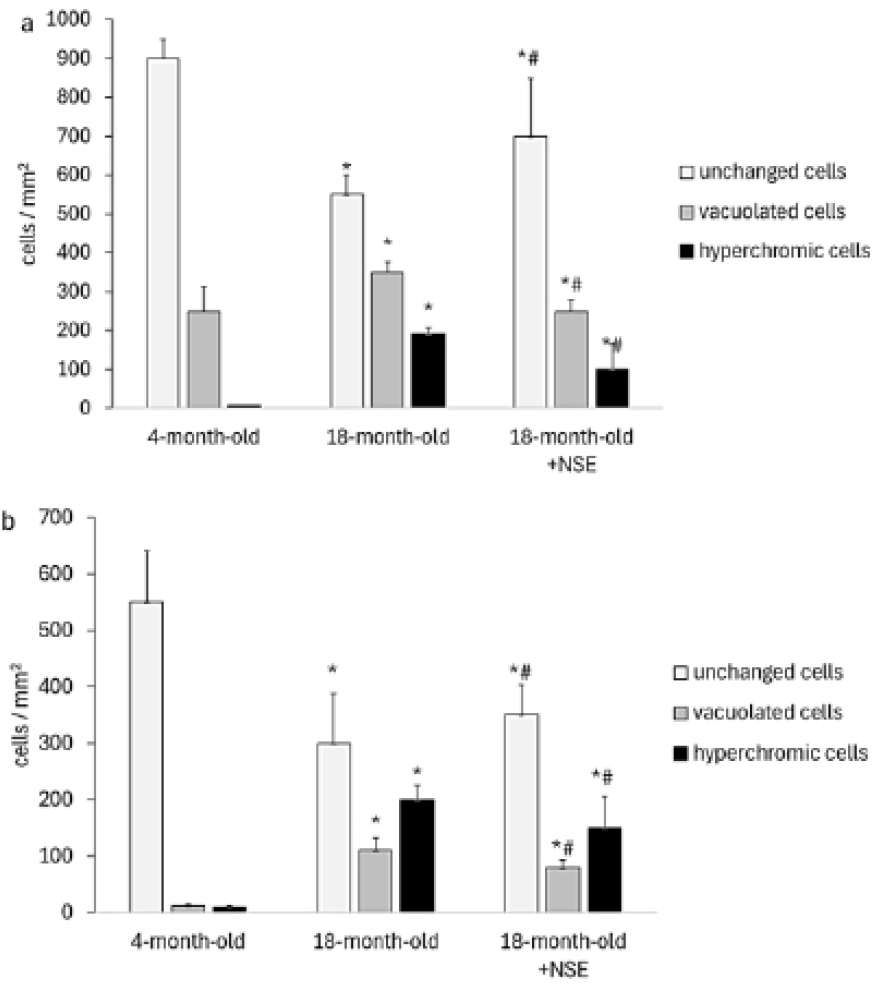
Number of unchanged (normal), vacuolated, and hyperchromic cells in the hippocampus (a) and frontal cortex (b) of rats. NSE, N-stearoylethanolamine; +NSE, treatment with water suspension of N-stearoylethanolamine at a dose of 50 mg/kg of body weight daily for 10 days; n=18-20; The numerical density of neurons was calculated as the sum of neurons per test area of 320x240 μm2, expressed in the number of cells per mm2. Data expressed as Mean ± SD. The Shapiro-Wilk test was used to assess the normality of the data. One-way ANOVA followed by a post hoc Tukey honestly significant difference (HSD) test was used to assess differences in all measurement indices between groups; *represents a statistically significant difference between 4-month-old and 18-month-old rat groups at the significance level of 0.05; # represents a statistically significant difference between the 18-month-old rats group and the same age group after NSE administration at the significance level of 0.05.
Fig. (7) shows that in old 18-month-old rats, the number of unchanged (normal) neurons in both the hippocampus (from 900±35 cells to 550±50 cells, Fig. (7a) and in the frontal cortex (from 550±90 to 300±88, Fig. (7b) significantly decreases. At the same time, the number of vacuolated and hyperchromic cells in both brain structures significantly increases. Thus, the number of vacuolated cells in the hippocampus significantly increased from 250±63 in 4-month-old rats to 350±25 cells in 18-month-old rats, and in the frontal cortex from 12±3 in 4-month-old rats to 110±22 cells in 18-month-old rats. The number of hyperchromic neurons in the hippocampus of 18-month-old rats was also significantly increased compared to their number in 4-month-old animals (190±18 cells and 6±1 cells, respectively). In the frontal cortex of 18-month-old rats, there is also an increase in the number of hyperchromic neurons (from 10±2 cells in 4-month-old rats to 200±26 cells in old rats).
In the treatment of 18-month-old NSE rats, the number of unchanged (normal) neurons in the hippocampus significantly increased from 550±50 cells in untreated old rats to 700±150 cells. Treatment with NSE also resulted in a reduction in the number of both vacuolized neurons (from 350±25 cells to 250±29 cells) and hyperchromic neurons (from 190±18 cells to 100±66 cells) in the hippocampus of 18-month-old rats. In the frontal cortex, NSE treatment also showed a significant reduction in the number of vacuolated cells (from 110±22 cells to 80±13 cells) and hyperchromic cells (from 200±26 cells to 150±55 cells).
4. DISCUSSION
4.1. The effect of NSE on Memory and Exploratory Activity
Various behavioral tests are widely used to confirm changes in cognitive function and memory [31-33]. Data from the literature show that aged rats have a decline in cognitive abilities [34]. Our study also found (Fig. 1) a decrease in investigated cognitive functions in old rats compared to their indicators in young animals. NSE administration had a positive effect on cognitive activity in old rats.
4.2. The Effect of NSE on the Lipid Composition of the Brain's Frontal Cortex and Hippocampus
The literature indicates that plasmalogen content in the human brain undergoes a significant increase during postnatal development, particularly during the formation of myelination processes, reaching peak values at 30 [35]. This data underscores the crucial role of plasmalogens in these developmental processes [36, 37], which is a key aspect of our research.
Given the critical role of plasmalogens in ensuring the physicochemical properties of neuronal cell membranes [38, 39], a decrease in their content in brain structures during aging can be considered a prerequisite for age-related brain disorders. This decline, in the long run, could lead to the development of neurodegenerative diseases, highlighting the potential impact of our research.
In our studies, the content of PtdEth and PlsEth in the frontal cortex of the brain of rats showed statistically significant changes during aging (Fig. 2). These data are consistent with the literature that shows that plasmalogen PlsEth levels decrease during normal aging and in some pathological conditions [40]. Our research demonstrates normalizing in PtdEth and PlsEth levels in the frontal cortex and changes in PtdEth and PlsEth levels in the hippocampus of old rats after NSE administration (Fig. 2).
The normalization of PtdEth and PlsEth amounts in the cortex of old rats under the influence of NSE can be considered one of the mechanisms of NSE neuroprotective action in aging. We do not know about the biochemical mechanisms of NSE's influence on the content of PtdEth and PlsEth in the brain, and the elucidation of these mechanisms requires additional research. It can be assumed that the so-called “entourage effect” of NSE may take place here [41, 42]. This effect is that the administration of exogenous NSE leads to a decrease in the activity of fatty acid amide hydrolase (FAAH), as a result of which the amount of other NAEs (e.g., anandamide or N-palmitoylethanolamine) may increase. It is known that some saturated NAEs can influence FAAH expression [43, 44]; among them, there are also high-affinity PPARα ligands [45]), which indicates the possibility of these compounds participating in memory formation processes. These other NAEs can activate PPARα. PPARα is known to regulate lipid metabolism and, in particular, can affect the fatty acid composition and amount of various phospholipids [46-48].
The literature has described a decrease in the content of the main phospholipids in the structures of the human brain during aging, with the most significant changes in phospholipid composition occurring in the frontal cortex and hippocampus [11, 49]. We obtained similar results in our research (Fig. 3). (3) shows that during aging, the PtdCho, Ptdlns, and PtdSer content in the frontal cortex and DPG, Ptdlns, and PtdSer in the hippocampus decreased. At the same time, we found an increase in the levels of DPG, CerPCho, LysoPtdCho, and PA in the frontal cortex and PtdEth and CerPCho in the hippocampus. The administration of NSE to old rats facilitated normalization of the content of DPG and PA in the frontal cortex and PtdCho, LysoPtdCho, DPG, and PA in the hippocampus.
It should be noted that we obtained a similar effect regarding the influence of NSE on the content of phospholipids earlier on various models of pathological conditions in animals. Moreover, the changes caused by NSE were, as a rule, aimed at eliminating the PL imbalance caused by the simulation of a particular pathological condition. At the same time, the administration of NSE to normal (control) animals did not cause probable changes in PL composition [50, 51]. Cholesterol in the mammalian brain is known to play a crucial structural and functional role [52, 53]. Various changes in the composition of brain lipids, including a decrease in cholesterol levels, are known to be associated with normal and pathological aging processes [54, 55]. In our study, the cholesterol content in the brain of 18-month-old rats was significantly lower than that of 4-month-old rats (Fig. 4). Cholesterol plays a crucial role in neurogenesis and synaptogenesis, and the observed age-related decline in cholesterol levels could lead to synaptic loss, impaired synaptic plasticity, and potentially contribute to the development of various neurodegenerative and cognitive disorders [48, 56-58].
Our study also indicates a decline in memory and exploration activity in old rats compared to young animals. The administration of NSE appears to contribute to an increase in cholesterol content in the hippocampus while not affecting its amount in the frontal cortex of the brain of old rats (Fig. 4). Importantly, this increase in cholesterol content is associated with a positive effect on memory and exploration activity in old rats (Fig. 1), offering encouragement for potential interventions in age-related cognitive decline. When NSE was administered to 18-month-old rats, cholesterol levels in the frontal cortex and hippocampus increased compared to those of untreated 18-month-old rats (p<0.05). However, it did not reach the cholesterol levels in the brains of 4-month-old rats (Fig. 4). The increase in cholesterol levels in the frontal cortex and hippocampus with NSE administration correlates with an increase in DI (r=0.79 for the frontal cortex, r=0.80 for the hippocampus) and the “number of head tilts” (r=0.50 for the frontal cortex, r=0.55 for the hippocampus) (Fig. 1).
Age-related cholesterol decreases can be caused by age-related increases in neuronal CYP46A1 and changes in the expression of cholesterol biosynthesis genes in the brain [59, 60], age-related increases in HMGCR, ApoE, and CYP46 mRNA levels [61], and, as a result, ATP-binding cassette transporter 1 (ABCA1) deficiency [62]. Different transcription and post-transcription factors, including PPARs, are known to modulate ABCA1 expression [63]. Some studies describe that PPARγ activators induce the expression of the gene encoding ABCA1 [64, 65]. Treatment with low-dose PPARγ agonists counteracts metabolic disorders [66], cognitive dysfunctions [67], and prolongs lifespan in aging mice [68]. Previously, we demonstrated that NSE is a PPARγ receptor agonist, causing changes in the expression of PPARγ-dependent genes [69, 70]. In our opinion, this may explain the effect of NSE on the cholesterol content in the hippocampus of old rats under the conditions of our experiment. However, such a hypothesis requires additional research. Thus, the results demonstrate that administering NSEs to old rats reduces age-related cholesterol loss in the brain.
4.3. Effects of NSE on the Choline and Acetylcholine Content and AChE Activity in the Blood Plasma and in the Brain's Frontal Cortex and Hippocampus
It is known that acetylcholine deficiency is the basis of age-related cognitive disorders of both neurodegenerative and vascular nature, which is an integral component of the pathogenesis of most neurodegenerative diseases. Dysfunctions of the cholinergic system, such as decreased choline absorption, increased AChE activity, decreased acetylcholine synthesis, decreased activity, and degeneration of cholinergic neurons in specific brain structures, are known to be associated with impaired cognitive function in people with Alzheimer's disease [71-74]. With aging, changes in AChE activity have been found to occur in the brain and blood plasma of rats [75] and various bird species [76].
Our research findings on AChE activity in the hippocampus, frontal cerebral cortex, and blood plasma of rats of different age groups, presented in Fig. (5), are significant in understanding the relationship between AChE activity, age, and cognitive function. Fig. (5 a-c) demonstrates that in the hippocampus and frontal cortex of old rats' brains, compared with young rats, the activity of AChE is compensatorily reduced. At the same time, the result showed statistically significant growth of AChE activity in the blood plasma of 18-month-old rats compared to the 4-month-old animals’ group. Administration of NSE to old rats increased the compensatory reduction of AChE activity (Fig. (5 a-c)), which contributes to the restoration of acetylcholine levels both in the hippocampus and in the frontal cortex of the brain. NSE administration positively affected blood plasma AChE activity in old rats, but its level remained higher than in young rats. In our previous studies in a rat model of scopolamine-induced cholinergic deficiency, we found that NSE can prevent increased AChE activity in the hippocampus and frontal cortex of rats [17]. In vitro studies also showed that the anti-inflammatory effect of NSE can be mediated through the activation of α7 nicotinic acetylcholine receptors [16] and PPARγ/NF-κB signaling pathways [65]. The literature describes the activation of AChE under the influence of pro-inflammatory factors and mediators [77]. The regulation of AChE activity by various anti-inflammatory drugs [78-80], both steroidal and non-steroidal in nature (dexamethasone, celecoxib, indomethacin, piroxicam, diclofenac, etc.) [81, 82], has also been shown to be more effective inhibitors of the enzyme compared to steroid ones [83]. In our studies, a decrease in AChE activity in the brain structures of old rats was found when using NSE (Fig. (5a-c). Given the anti-inflammatory effect of NSE, it can be assumed that reducing the level of inflammatory processes in the rat brain due to its action will also contribute to a decrease in AChE activity.
According to the literature, increased oxidative stress, decreased antioxidant enzyme activity, altered AChE activity, and decreased levels of biogenic amines in the brains of old rats correlate with memory impairment [71]. In our study, a moderate positive correlation was found between DI values and AChE activity in both the cerebral cortex (r = 0.55) and the hippocampus (r = 0.53). Head-dip count values were weakly correlated with AChEactivity in the frontal cortex (r=0.21) and in the hippocampus (r=0.16). Thus, it can be assumed that NSE, by influencing AChE activity, contributes to the improvement of memory in old rats.
Among all neurotransmitters, acetylcholine (of which choline is a precursor) plays an essential role in the formation of all types of memory. Basal cholinergic neurons of the forebrain, which exert a cholinergic effect on the cerebral cortex and hippocampus, undergo degenerative changes during the aging process. It is the cholinergic dysfunction that occurs in this way that is thought to be the cause of progressive memory impairment in the elderly [84-86].
The choline content in the brain is known to be crucial for optimal cognitive function [80]. In our studies, 18-month-old rats had increased choline content compared to a group of 4-month-old rats. Administration of NSE to old rats reduced choline levels in the frontal cortex and hippocampus, but the amount remained significantly higher than in 4-month-old rats (Fig. (5 d-e)).
The data presented in Fig. (5 f,g) show that aging is accompanied by a significant decrease in the level of acetylcholine in the frontal cortex and hippocampus of rats compared to its level in young animals. Administration of NSE caused an increase in acetylcholine in the hippocampus. At the same time, in the frontal cortex of the brain, the level of acetylcholine was at the level found in 4-month-old rats.
Previously, we showed the inhibitory effect of NSE on AChE activity in the hippocampus and frontal cortex of the rat brain when modeling scopolamine-induced cholinergic deficiency [17]. Recent research suggests the possibility of regulatory interactions between the cholinergic system of rodents and the endocannabinoid regulatory system [87]. The regulatory effect of oleoylethanolamine (NAE 18:1) on the activity of choline acetyltransferase when modeling scopolamine-induced cognitive disorders in mice [88] is also known. We hypothesize that the effect of endocannabinoid NSE on choline (Fig. d, e) and acetylcholine (Fig. 5f, g) in the frontal cortex and hippocampus of the brain of old rats is due to its modulating effect on AChE activity. Thus, the correlation coefficient r = 0.73 between AChE activity and acetylcholine content in the hippocampus; r = - 0.17 between the activity of AChE and the content of acetylcholine in the frontal cortex. r = -0.83 between plasma AChE activity and hippocampal acetylcholine content; r = -0.61 between the activity of AChE in plasma and the content of acetylcholine in the frontal cortex. However, this hypothesis requires more research in the future.
4.4. Effect of NSE on Age-related Morphological Changes in Rat Brain Tissue
There is evidence that with aging, the brain undergoes various morphofunctional changes that are often accompanied by the development of impairments of cognitive and sensorimotor functions [89-92]. Our study also found a decrease in cognitive functions investigated in old male rats compared to those in young animals. NSE administration positively affected cognitive activity in old rats (Fig. 1). Consequently, based on the obtained data on the NSE effect on certain biochemical characteristics and investigated cognitive functions in old rats, it was also reasonable to investigate the influence of NSE on brain tissue morphofunctional changes in aged rats.
Analysis of the cytoarchitectonics of the hippocampal CA1 region (Fig. 6a) revealed 3 conditional types of neurons: unchanged (intensely stained, with an intact cell membrane, with a gently stained blue-violet cytoplasm and clearly defined nuclei), hyperchromic (with an intensely stained cytoplasm that merges with the color of the cell the membrane, which can often be damaged or deformed, it is very hard to distinguish the nucleus) and neurons with signs of edema (“vacuolated”, the cytoplasm is almost not stained, the cell membrane is damaged, the nucleus is almost invisible or absent). The latter often bordered the zones of pericapillary and pericellular edema. In the hippocampal CA1 region of old rats, compared to young animals, the percentage of hyperchromic and vacuolated neurons, as well as vacuolated gliocytes (a significant part of which may be in a state of apoptosis), rose significantly. Also, increased pericellular and pericapillary swelling is observed in old rats. In the hippocampal CA1 region of old rats, compared to young animals, the count of hyperchromic and vacuolated neurons significantly increased, with a simultaneous decrease in the number of normal neurons. Also, pericellular and pericapillary swelling was observed in old rats.
Administration of NSE to old rats resulted in a significant increase in the number of normal neurons in the hippocampus and a decrease in the number of vacuolated neurons with signs of edema and hyperchromic cells (Fig. 7a). Such a change in the number of normal, vacuolated, and hyperchromic cells in the hippocampus for all animal groups correlated with changes in the value of the DI index (r = 0.71, -0.60, -0.80, respectively). The correlation coefficient between changes in the head-dip-count test and the number of normal, vacuolated, and hyperchromic cells was 0.63, -0.66, and -0.72, respectively. These findings indicate a reduction in the manifestations of age-related metabolic changes in these brain cells under the influence of NSE.
In the sensorimotor part of the frontal cortex of old rats, we observed a significant decrease in the number of unchanged neurons and an increase in the number of vacuolated and hyperchromic neurons compared to young rats (Fig. 7b). In addition, there was an increase in pericellular and pericapillary edema (Fig. 6b). Administration of NSE to old rats resulted in a significant increase in the number of normal unchanged neurons and a decrease in the number of vacuolated and hyperchromic neurons. Such a change in the number of normal, vacuolated, and hyperchromic cells in the frontal cortex for all animal groups is moderately correlated with changes in the DI index value (r = 0.71, -0.80, -0.78, respectively). The correlation coefficient between changes in the head-dip-count test and the number of cells of different types in the frontal cortex was 0.46, -0.68, and -0.62, respectively.
The described results suggest a potential reduction in the manifestations of age-related metabolic changes in the hippocampus and frontal cortex of 18-month-old rats under the influence of the NSE, which also contributes to improved memory and sensorimotor activity.
5. STUDY LIMITATIONS
While the present study provides valuable insights into the neuroprotective and antiamnestic effects of NSE in age-related changes in the rat brain, several limitations should be acknowledged. First, the sample size was limited to a specific age group and animal model, which may restrict the generalizability of the findings to other species or broader age ranges. The exclusive use of male rats may also introduce gender-based bias, as sex differences in neurodegenerative processes and response to treatment have been reported in previous literature. Additionally, although biochemical, behavioral, and morphological endpoints were assessed, the study did not account for possible confounding factors such as environmental stressors or individual variability in baseline cognitive function.
Another constraint is the short duration of NSE administration and follow-up, which may not fully capture the long-term efficacy and safety profile of the compound. The mechanistic pathways underlying NSE’s effects, particularly its interaction with the cholinergic and endocannabinoid systems, remain incompletely understood and warrant further investigation using molecular and genetic approaches. Finally, the lack of direct comparison with other neuroprotective agents limits the ability to contextualize NSE’s therapeutic potential within the broader landscape of interventions for age-related neurodegeneration.
Future studies should aim to address these limitations by including larger and more diverse cohorts, exploring longer-term treatment outcomes, and employing advanced analytical techniques to elucidate the molecular mechanisms involved. Comparative studies with established neuroprotective compounds would also strengthen the evidence base and clarify the unique benefits of NSE. These steps are essential for translating preclinical findings into effective clinical strategies for managing age-related cognitive decline.
CONCLUSION
Thus, the results of this study suggest that NSE substantially improves selected behavioral responses and biochemical parameters in the brains of aged male rats. Specifically, NSE treatment enhances cognitive functions and reduces the severity of morphofunctional disorders in brain structures, including a reduction in signs of age-related neuronal degeneration. Our data confirm the involvement of NSE, as a component of the endocannabinoid system, in the regulation of the cholinergic system, and highlight the potential for further study of this compound in mitigating age-related neurodegenerative processes.
LIST OF ABBREVIATIONS
| AChE | = Acetylcholinesterase |
| ApoE | = Apolipoprotein E |
| CB | = Cannabinoid receptors |
| CerPCho | = Sphingomyelin |
| CYP46 | = Gene that codes for the enzyme cholesterol 24-hydroxylase |
| DPG | = Diphosphatidylglycerol |
| DTNB | = Dithio p-nitrobenzoic acid |
| FAAH | = Fatty acid amidohydrolase |
| HMGCR | = 3-hydroxy-3-methyl-glutaryl-coenzyme A reductase |
| LysoPtdCho | = lysophosphatidylcholine |
| NAE | = N-acylethanolamines |
| NOR | = “Novel object recognition” test |
| NSE | = N-stearoylethanolamine (NAE 18:0) |
| PA | = Phosphatidic acid |
| Pi | = Inorganic phosphorus |
| PL | = Phospholipids |
| PPAR | = Peroxisome proliferator–activated receptors |
| PtdCho | = Phosphatidylcholine |
| PtdEtn | = Phosphatidylethanolamine |
| PtdGro | = Phosphatidylglycerol |
| Ptdlns | = Phosphatidylinositol |
| PtdSer | = Phosphatidylserine |
| SD | = Standard deviation |
| TNB | = 5-mercapto-nitrobenzoic acid |
AUTHOR’S CONTRIBUTIONS
The authors confirm their contribution to the paper as follows: study conception and design: HK; data collection: OM, VK, TKR, OT, OZ; Data analysis and interpretation: TH, SM; Conceptualization: NH; Writing the paper: AB. All authors reviewed the results and approved the final version of the manuscript.
ETHICS APPROVAL AND CONSENT TO PARTICIPATE
The study was approved by the Animal Care and Use Committee of the Palladin Institute of Biochemistry of the NAS of Ukraine (protocol No 8, 05.05.2021).
HUMAN AND ANIMAL RIGHTS
All experiments involving animals were conducted following established bioethical principles and international regulations for the use of laboratory animals, including EU Directive 2010/63/EU, the European Convention for the Protection of Vertebrate Animals Used for Experimental and Other Scientific Purposes (Strasbourg, 1986), and the Ukrainian Guidelines for Bioethical Assessment of Preclinical and Other Scientific Research on Animals (Kyiv, 2006).
This study adheres to internationally accepted standards for animal research, following the 3Rs principle. The ARRIVE guidelines were employed for reporting experiments involving live animals, promoting ethical research practices.
FUNDING
This research was financially supported by the National Academy of Sciences of Ukraine. The specific funding was provided under grant numbers 0117U004344 and 0119U002510, as well as through projects N 0119U002510 and N 0117U004344. These grants and projects played a crucial role in enabling the various aspects of the study.
ACKNOWLEDGEMENTS
Declared none.
References
[PubMed Link]
[PubMed Link]
[PubMed Link]
[PubMed Link]
[PubMed Link]
[PubMed Link]
[PubMed Link]
[PubMed Link]
[PubMed Link]
[PubMed Link]
[PubMed Link]
[PubMed Link]
[PubMed Link]
[PubMed Link]
[PubMed Link]
[PubMed Link]
[PubMed Link]
[PubMed Link]
[PubMed Link]
[PubMed Link]
[PubMed Link]
[PubMed Link]
[PubMed Link]
[PubMed Link]
[PubMed Link]
[PubMed Link]
[PubMed Link]
[PubMed Link]
[PubMed Link]
[PubMed Link]
[PubMed Link]
[PubMed Link]
[PubMed Link]
[PubMed Link]
[PubMed Link]
[PubMed Link]
[PubMed Link]
[PubMed Link]
[PubMed Link]
[PubMed Link]
[PubMed Link]
[PubMed Link]
[PubMed Link]
[PubMed Link]
[PubMed Link]
[PubMed Link]
[PubMed Link]
[PubMed Link]
[PubMed Link]
[PubMed Link]
[PubMed Link]
[PubMed Link]
[PubMed Link]
[PubMed Link]
[PubMed Link]
[PubMed Link]
[PubMed Link]
[PubMed Link]
[PubMed Link]
[PubMed Link]
[PubMed Link]
[PubMed Link]
[PubMed Link]
[PubMed Link]
[PubMed Link]
[PubMed Link]
[PubMed Link]
[PubMed Link]
[PubMed Link]
[PubMed Link]
[PubMed Link]
[PubMed Link]
[PubMed Link]
[PubMed Link]
[PubMed Link]
[PubMed Link]